23 ENTRIES · 9 WITH FIGURES FROM THE PAPERS

PUBLICATIONS

Every entry below carries its verified venue, year and DOI or preprint link. Where a paper has a figure I made, that figure is on the card — nothing here is an illustration of research that does not exist.

SHOWING 23 / 23

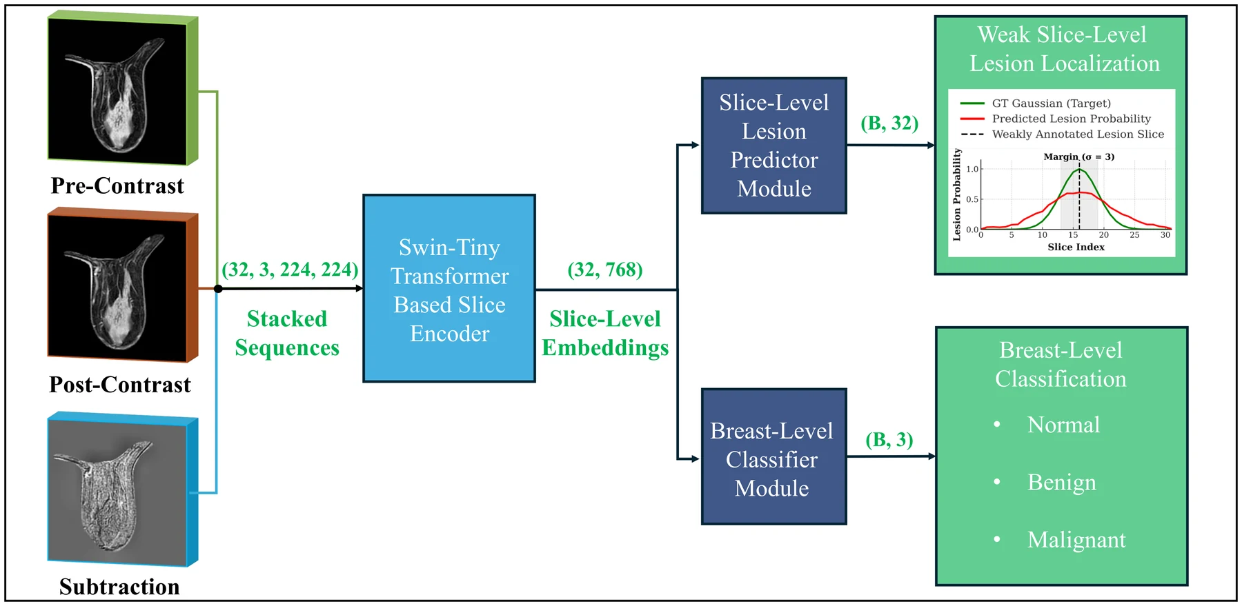
BE-WISE architecture: pre-contrast, post-contrast and subtraction MRI volumes stacked into a (32, 3, 224, 224) tensor, encoded by a shared Swin-Tiny slice encoder into (32, 768) embeddings, then split into a slice-level lesion predictor producing a per-slice probability curve and a breast-level classifier producing normal, benign or malignant

Bildverarbeitung für die Medizin (BVM) 2026, 10–17

BE-WISE: Breast MRI Evaluation with Weakly-Informed Slice-Level Explanation

Published

Breast MRI models usually need lesions outlined voxel by voxel, which does not scale. BE-WISE asks how far one annotated slice per exam gets you. Pre-contrast, post-contrast and subtraction volumes are stacked and encoded slice by slice with a shared Swin-Tiny transformer. One branch predicts a per-slice lesion probability against a Gaussian target centred on the radiologist's slice; the other pools the same embeddings by attention into a normal / benign / malignant decision. On the multicenter ODELIA dataset this reached test AUC 0.8683 and an ODELIA score of 0.7098, against 0.7968 and 0.5695 for a medical slice transformer baseline.
Explainable AIMedical ImagingDetection / Localization
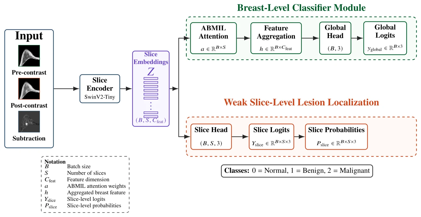
BE-WISER architecture: pre-contrast, post-contrast and subtraction inputs go through a SwinV2-Tiny slice encoder into slice embeddings, which split into a breast-level classifier module with ABMIL attention, feature aggregation, global head and global logits, and a weak slice-level lesion localization branch with a slice head, slice logits and per-slice class probabilities over normal, benign and malignant

Research Square preprint (in review)

BE-WISER: Class-Aware Weak Slice-Level Supervision for Breast MRI

Preprint · in review

BE-WISE could say which slice contained something; BE-WISER says what that something is. Each slice inside the Gaussian region around the radiologist's marked slice inherits the exam's class — benign or malignant — while the rest are labelled normal, so the slice head becomes three-class instead of binary. On the latest multicenter ODELIA release (926 / 230 / 292 examinations from six institutions) this reached breast-level AUC 0.864 and an ODELIA score of 0.699, against 0.807 and 0.591 for a breast-level baseline. Localization also improved: Hit@1 rose from 0.554 to 0.632 and the mean slice distance fell from 2.838 to 2.505 slices.

Explainable AIDetection / LocalizationMedical Imaging
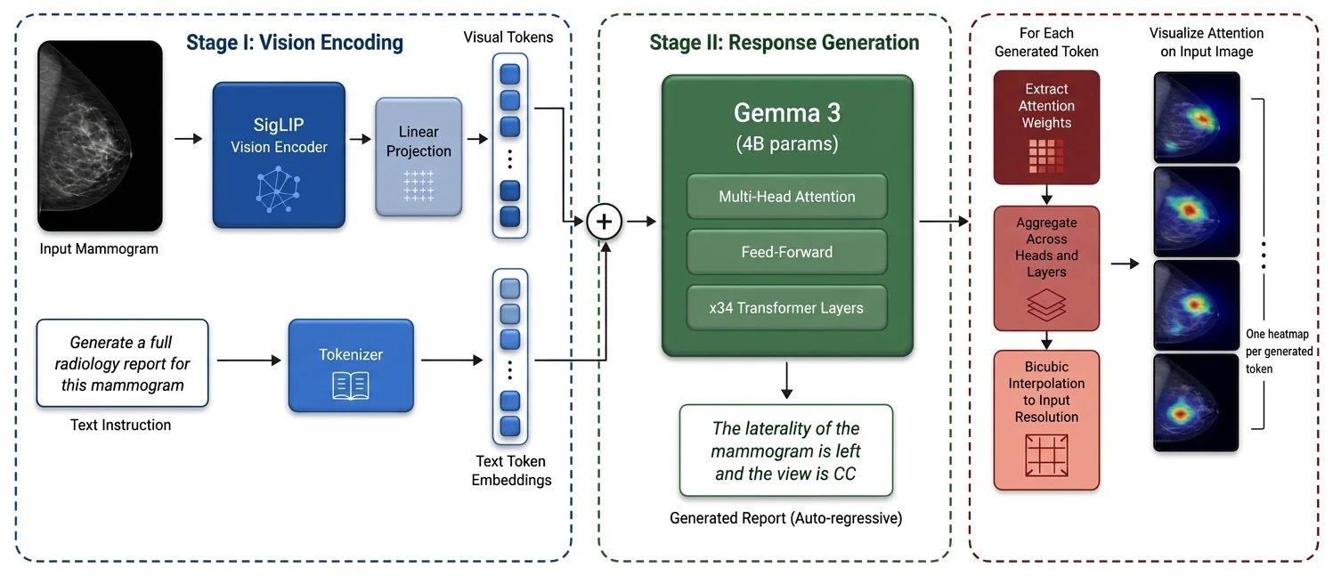
Three-stage architecture: an input mammogram through a SigLIP vision encoder and linear projection into visual tokens, a text instruction through a tokenizer into text token embeddings, both combined into a Gemma 3 4B model with 34 transformer layers generating a report autoregressively, then attention weights extracted per token, aggregated across heads and layers and bicubically interpolated to produce one heatmap per generated token

Manuscript in preparation — no venue yet

Explainable Vision-Language Models for Mammography Report Generation

Manuscript in preparation · not peer reviewed

This is the follow-up to MammoBLIP, and it asks a different question: not how good the report is, but whether you can see where each word came from. Domain-adapted SigLIP vision encoders are paired with 4B-parameter LLMs (Gemma 3 and MedGemma), and attention is aggregated across heads and layers to produce one heatmap per generated token. The best of six configurations reached ROUGE-1 0.877 and ROUGE-2 0.843, up 0.127 and 0.143 on MammoBLIP. The more interesting finding is negative: attention often lands on the right microcalcification cluster while the diagnosis is still wrong.

Vision-Language ModelsLLM / GenAIExplainable AI
conference 2026 Vision-Language Models · Foundation Models

European Congress of Radiology (ECR) 2026, scientific exhibit C-15780

Lesion-Aware AI for Mammography: Multi-Dataset Pretraining with ROI-Guided Contrastive Learning and Clinical Image Retrieval

Accepted

Global contrastive pretraining lets a mammography model align on breast density or view rather than on the lesion, because those cues are enough to match an image to its report. This work adds region-of-interest guidance to multi-dataset contrastive pretraining, so localized pathology has to contribute to the representation, and then tests the embeddings on clinical image retrieval — finding visually and diagnostically similar prior cases. Accepted as a scientific exhibit at ECR 2026; quantitative results are not yet publicly available.

Vision-Language ModelsFoundation ModelsMedical Imaging
conference 2026 Medical Imaging · Clinical Validation

Bildverarbeitung für die Medizin (BVM) 2026, 427–434

Opportunistic Breast Cancer Risk Stratification From Low-dose Chest CT Using Multiple Instance Learning

Published

Low-dose chest CT is already acquired at scale for lung screening, and it happens to include breast tissue. This proof-of-concept study applies multiple instance learning to that existing data for opportunistic breast cancer risk stratification, stratified against BI-RADS 3–6 categories on a cohort of 60 patients. The individual-breast model reached 72.7% accuracy with AUC 0.85, and the bilateral model 75.0% accuracy with AUC 0.87. The authors describe it as the first comprehensive proof-of-concept of its kind. I co-supervised the master's thesis this paper grew out of and co-authored the result.

Medical ImagingClinical Validation
journal 2026 Vision-Language Models · Explainable AI

Scientific Reports, 16, article 14688

PatchCLIP enables region specific contrastive health record and image joint training with patch embedding loss

Published · open access (CC BY 4.0)

Contrastive image-text models in radiology align a whole image with a whole report, so their explanations have to be reverse-engineered with saliency methods afterwards. PatchCLIP instead trains a patch-level contrastive objective, so localization is part of the model rather than a post-hoc interpretation. At inference it emits patch-wise prediction maps as well as a global score. Across eight chest X-ray abnormality detection tasks the paper reports the best performance of the methods it compares against, and substantially fewer false positives at comparable sensitivity than saliency-map baselines. Code is released publicly by Siemens Healthineers. I contributed as a co-author.

Vision-Language ModelsExplainable AIDetection / Localization
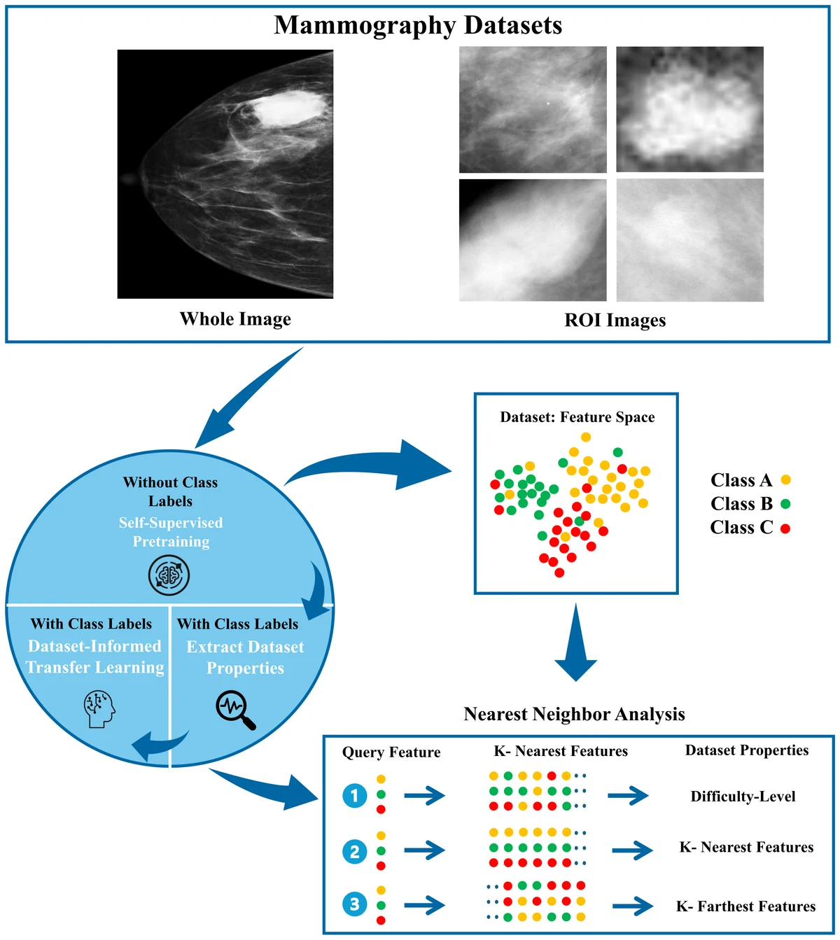
DITL framework diagram: whole-image and ROI mammography inputs feed a three-stage circle of self-supervised pretraining without labels, dataset property extraction with labels, and dataset-informed transfer learning; a feature-space scatter and a nearest-neighbour analysis panel show difficulty level, k-nearest and k-farthest features

arXiv preprint (accepted at the International Conference on Computer Vision and Image Processing, CVIP 2025 — in print)

Re-thinking Mammography Transfer Learning: The Dataset-Informed Transfer Learning (DITL) Framework for Breast Cancer Screening and Lesion Diagnosis

Preprint · CVIP 2025 in print

My earlier neighbourhood-based loss worked on one small dataset but did not scale: the difficulty weights were hand-set, the triplet margin was fixed, and the number of neighbours had to be tuned per dataset. DITL replaces all three with quantities the dataset supplies itself — difficulty from nearest-neighbour label purity in a self-supervised feature space, a learnable margin, and k set to the square root of the dataset size. Neighbour statistics are precomputed offline, so training cost barely changes. Across VinDr-Mammo, CDD-CESM and two CBIS-DDSM subsets it beats cross-entropy, focal loss and my own previous best, reaching macro F1 0.609 on VinDr-Mammo breast density.

Foundation ModelsSelf-Supervised LearningMedical Imaging
conference 2025 Clinical Validation · Medical Imaging

European Congress of Radiology (ECR) 2025, EPOS scientific exhibit C-21046

AI-Assisted Detection of Malignant Breast Cancer in High Breast Density Cohorts: A Retrospective Comparative Study

Published (e-poster)

This follow-up to the ECR 2024 study focuses on the subgroup where mammography is weakest: dense breasts with a suspicious finding. A retrospective 12-month Hanoi Medical University Hospital cohort of 443 patients (BI-RADS 4, 5 and selected 3) was assessed against biopsy-confirmed outcomes — 136 negative cases, 292 with unilateral lesions of which 138 were malignant, and 15 bilateral cases with 3 malignant. Inter-rater agreement between readers was ICC 0.718–0.780 (p < 0.001). The commercial AI independently identified 97% of positive patients correctly, while classifying 21% of biopsied patients as low risk.

Clinical ValidationMedical Imaging
conference 2025 Clinical Validation · Detection / Localization

European Congress of Radiology (ECR) 2025, EPOS scientific exhibit C-21110

Assessment of AI Performance in Detecting Breast Lesions on MRI: A Retrospective Study in a Chinese Cohort

Published (e-poster)

A breast MRI AI prototype developed in Europe was evaluated retrospectively on an external Chinese cohort — the kind of test that decides whether a model generalizes or merely fits its development population. 200 cases were sampled from a 4,000-patient West China Hospital breast MRI cohort acquired between November 2017 and August 2023, using multi-sequence 3T MRI with expert lesion annotations. Case-level sensitivity was 0.906, with a median of 0 false positives per case, an average of 0.635, and a maximum of 7 in a single case. This work came out of a guest research stay at the hospital.

Clinical ValidationDetection / LocalizationMedical Imaging
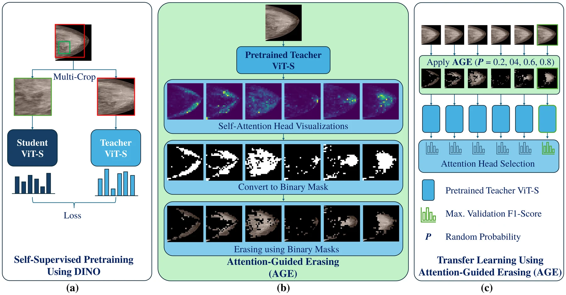
Three-panel diagram: DINO self-supervised pretraining with a student and teacher ViT-S, conversion of six attention heads into binary masks and erased images, and transfer learning with attention-head selection by validation F1

International Journal of Computer Assisted Radiology and Surgery, 20(3), 433–440

Attention-guided erasing for enhanced transfer learning in breast abnormality classification

Published · open access (CC BY 4.0)

Transfer learning helps when medical imaging datasets are small, but common augmentations such as random erasing are blind to anatomy and can delete the very evidence a classifier needs. This paper uses the self-attention maps of a DINO-pretrained Vision Transformer — trained on the target cohort without any labels — as the erasing policy: keep what the model attends to, erase the surrounding background at random. The attention head is chosen per task by validation F1. Across five mammography classification tasks on VinDr-Mammo, CDD-CESM and CBIS-DDSM, mean macro F1 improved significantly in four. The fifth, mass ROIs, barely moved because the lesion already filled the crop.

Self-Supervised LearningMedical ImagingExplainable AI
conference 2025 Detection / Localization · Medical Imaging

Medical Image Computing and Computer Assisted Intervention (MICCAI) 2025, LNCS, 205–215

Exemplar Med-DETR: Toward Generalized and Robust Lesion Detection in Mammogram Images and Beyond

Published

Detectors trained on one mammography cohort tend to collapse on another, especially in dense breasts. Exemplar Med-DETR learns class-specific exemplar features inside the model and fuses them with image features through cross-attention, so detection is anchored to prototypes rather than to raw appearance. On the Vietnamese VinDr-Mammo cohort it reports mAP 0.70 for mass and 0.55 for calcification detection — a 16 percentage-point absolute improvement over the previous state of the art — and roughly a twofold gain on an out-of-distribution Chinese cohort. It also transfers to chest X-ray and angiography, improving by 4 and 7 points. I contributed as a co-author.

Detection / LocalizationMedical ImagingFoundation Models
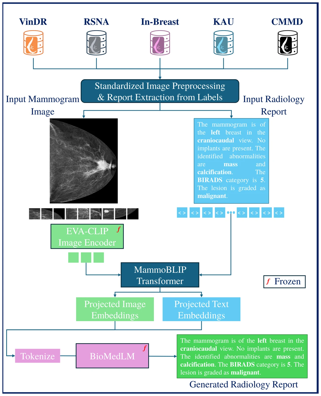
MammoBLIP pipeline: five public dataset sources feed a standardized preprocessing step; a mammogram goes through a frozen EVA-CLIP image encoder and the paired report through a tokenizer, both into the MammoBLIP transformer, whose projected embeddings drive a frozen BioMedLM to produce a generated radiology report

2025 IEEE Nuclear Science Symposium, Medical Imaging Conference and Room Temperature Semiconductor Detector Conference (NSS/MIC/RTSD), Yokohama

MammoBLIP: End-to-End Mammography Report Generation with Vision-Language Models and Public Multi-Institutional Datasets

Published

Most breast imaging AI outputs a single number. This work asks whether a model can instead write the structured report — laterality, view, abnormality type, BI-RADS category, malignancy grade — directly from a mammogram. Five public datasets were curated into one standardized 81,076-image corpus with reports derived from their labels. A frozen EVA-CLIP vision encoder and a frozen BioMedLM language model are joined by a small trained bridge transformer, so only the bridge and projection heads are learned. On 13,242 held-out report pairs the generated text reached BLEU 65.36, ROUGE-1 0.75, BERT-F1 0.88 and SBERT similarity 0.91, at roughly a seventh-grade reading level.

Vision-Language ModelsLLM / GenAIMedical Imaging
workshop 2025 Detection / Localization · Multimodal AI

Deep-Breath Workshop, MICCAI 2025, LNCS, 258–267

MM-DETR: Emulating the Diagnostic Clinical Workflow in Multi-view Multi-modal Mammography Mass Detection

Published

A radiologist never reads one mammographic projection in isolation — the craniocaudal and mediolateral oblique views are compared before a finding is called. Most detectors ignore that. MM-DETR adds bidirectional cross-attention between the paired views inside a DETR-style detector, so evidence in one projection can support or suppress a detection in the other. On VinDr-Mammo it reports mass-detection mAP 0.654 against 0.580 for Mammo-CLIP — 7.4 points higher in absolute terms, which the paper states as a 12.8% relative margin — and a 5.9% lower false-negative rate in the dense-breast (density C) cases where single-view reading struggles most. I co-supervised and co-authored this work.

Detection / LocalizationMultimodal AIMedical Imaging
preprint 2025 Foundation Models · Vision-Language Models

Journal submission — under internal review

Towards Foundational Models in Mammography: Leveraging Vision-Language Models for Radiological Reasoning

Under internal review · no results disclosed

A journal manuscript on vision-language foundation models for mammography, focused on whether image-text representation learning can support structured radiological reasoning rather than a single classification output. It is listed here for completeness of the record: the work is under internal review, no venue has been assigned, and no quantitative results are publicly available, so this page deliberately contains no numbers. The related and more developed strand of this line of work — token-level attention grounding of generated mammography reports — is written up separately on this site.

Foundation ModelsVision-Language Models
preprint 2025 Medical Imaging

Journal submission — under internal review

Towards Improving Mammography Quality: CNN Models for Skin Fold Positioning Error Classification

Under internal review · no results disclosed

A skin fold in a mammogram is a positioning error: it can obscure tissue, degrade the read, and force a repeat exposure. This work trains convolutional neural networks to detect that error automatically, so quality control happens at acquisition rather than at reporting. It comes out of the Siemens Healthineers part of my doctoral work with the mammography systems team. The manuscript is under internal review, no venue has been assigned, and no quantitative results are publicly available, so this page deliberately contains no numbers.

Medical Imaging
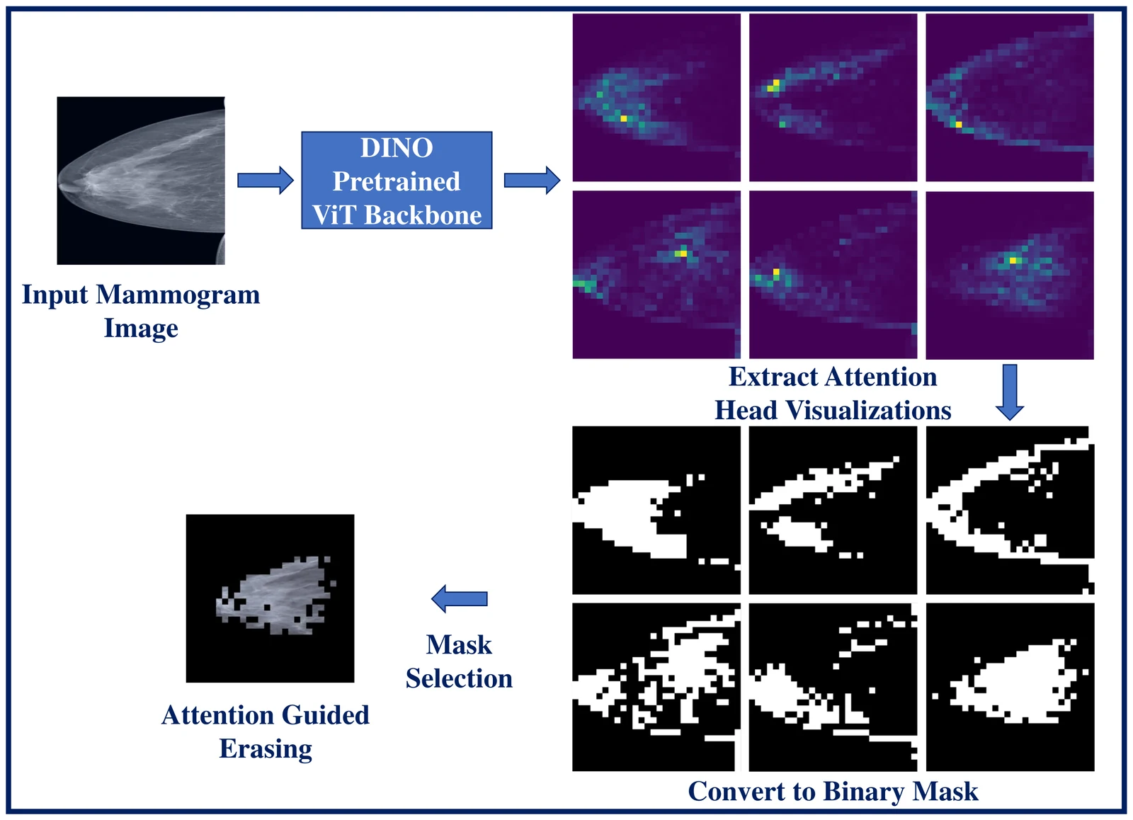
Method diagram: an input mammogram passes through a DINO-pretrained Vision Transformer backbone, producing six attention head visualizations, which are converted into six binary masks; after mask selection the result is a single attention-guided erased mammogram showing only the dense tissue region

Bildverarbeitung für die Medizin (BVM) 2024, Erlangen, 13–18

Attention-Guided Erasing: A Novel Augmentation Method for Enhancing Downstream Breast Density Classification

Published

Breast density affects how much a mammogram can hide, so automating BI-RADS density assessment matters — but the usual pipeline needs a separate segmentation network trained on radiologist annotations. This paper skips that. A Vision Transformer is pretrained with DINO on the target cohort with no labels, one of its attention heads is thresholded into a binary mask that roughly follows dense tissue, and everything outside is erased at random during fine-tuning. On the VinDr-Mammo test set of 4,000 images this reached mean macro F1 0.5910, against 0.5594 without erasing and 0.5691 with random erasing. The rarest class, density A, improved most.

Self-Supervised LearningMedical Imaging
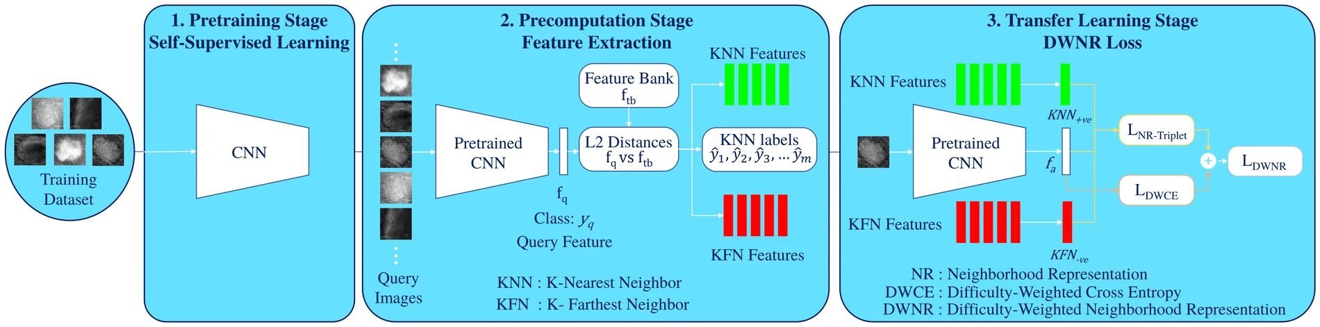
Three-stage horizontal diagram: pretraining stage with self-supervised learning on a CNN over CESM training patches; precomputation stage where query features are compared by L2 distance against a feature bank to yield k-nearest and k-farthest features and neighbour labels; transfer learning stage where those features feed a neighbourhood triplet loss and a difficulty-weighted cross-entropy that sum to the DWNR loss

SPIE Medical Imaging 2024: Computer-Aided Diagnosis, vol. 12927, 129270F

Enhancing downstream classification of breast abnormalities in contrast enhanced spectral mammography using a neighborhood representation loss

Published

Contrast-enhanced spectral mammography has too little public data for self-supervised learning to shine on its own. This work extracts features once after SimCLR pretraining, then uses each sample's nearest same-class and farthest different-class neighbours in that feature space to build the training loss: samples whose neighbours disagree with their own label are weighted as harder, and the same neighbours serve as triplet positives and negatives without a Siamese network. On the 116-ROI CDD-CESM test set this lifted three-class F1 from 0.8159 to 0.8413 and AUC from 0.9278 to 0.9350 over cross-entropy — the first reported ROI classification results on this dataset.

Self-Supervised LearningMultimodal AIMedical Imaging
conference 2024 Clinical Validation · Medical Imaging

European Congress of Radiology (ECR) 2024, EPOS scientific exhibit C-16214

Enhancing mammography screening sensitivity with AI-assistance: Evidence from a Vietnamese study cohort

Published (e-poster)

Mammographic sensitivity drops in dense breasts, and dense tissue is common in Vietnamese screening populations. This retrospective study compared radiologist reads with and without commercial AI support on a Hanoi Medical University Hospital cohort collected between November 2022 and April 2023, from a base of 1,119 eligible patients. The AI-assisted read reported AUC 75.2% (95% CI 75.0–75.5) and sensitivity 61.5% (95% CI 60.8–61.6), against AUC 73.2% and sensitivity 57.5% for the unassisted read — roughly a four-point absolute sensitivity gain. It is a retrospective e-poster study, not a prospective trial.

Clinical ValidationMedical Imaging
journal 2023 Medical Imaging · Clinical Validation

Scientific Reports, 13, article 21097

AUCReshaping: improved sensitivity at high-specificity

Published · open access (CC BY 4.0)

A screening AI is only ever used at one operating point — the one where false positives are acceptable — yet models are trained to optimize the whole ROC curve. AUCReshaping changes the loss so that positive cases misclassified near a chosen high-specificity threshold get extra weight, pulling sensitivity up exactly where it will be read off. Evaluated on chest X-ray (16,953 training images), mammography (20,000 images) and credit-card fraud detection (285,299 transactions), it reports sensitivity gains of 2 to 40 percentage points at high-specificity operating points depending on the dataset and the threshold chosen.

Medical ImagingClinical Validation
preprint 2023 Medical Imaging · Self-Supervised Learning

arXiv preprint 2301.09282

Classification of Luminal Subtypes in Full Mammogram Images Using Transfer Learning

Preprint

Molecular subtype is determined from tissue, so the question here is whether the mammogram itself carries any usable signal for it. A ResNet-18 was transferred from a breast abnormality classification task and fine-tuned on the public CMMD dataset for luminal versus non-luminal classification, using image-level labels only — no tumour outlines. It reached mean AUC 0.6688 and mean F1 0.6693 on the test set, a statistically significant improvement over the baseline (p < 0.0001). That is well above chance and well below anything clinically usable, which is the honest reading of the result.

Medical ImagingSelf-Supervised Learning
journal 2023 Medical Imaging

International Journal of Laboratory Hematology, 45(S3), 3–137 (ISLH XXXVI International Symposium abstract)

Unstained White Blood Cell Classification Using Deep Learning

Published (conference abstract)

White blood cell morphology is normally read after staining, which costs time and reagents. This study tested whether deep learning can classify unstained cells directly, across 14 classes, on a dataset of 339,885 unstained images from 233 patients split 70/17/13% at patient level. A two-stage pipeline reached mean weighted F1 of 90.33% with a ResNet-18 backbone and 91.77% with a transformer, a 1.44-point gain for the transformer. It is the one piece of my work outside radiology, and it is published as a conference abstract rather than a full paper. I contributed as a co-author.

Medical Imaging
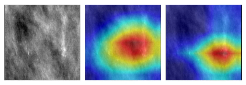
Three panels side by side: a grayscale mammography calcification patch, a Grad-CAM heatmap from the model trained without equalization showing a broad diffuse hot region, and a Grad-CAM heatmap from the model trained with random equalization at P equals 0.4 showing a tighter hot region over the bright calcification

Bildverarbeitung für die Medizin (BVM) 2022, Heidelberg, 173–178

Effect of Random Histogram Equalization on Breast Calcification Analysis Using Deep Learning

Published

Histogram equalization is usually applied to every mammogram before training. On small calcification crops that backfires — the bright spots that define the class get flattened into noise. This paper applies equalization only during training and only with probability P, so the model sees both versions. On the CBIS-DDSM calcification test set the three-class F1 rose from 0.6855 without equalization to 0.7071 at P = 0.4, while always equalizing dropped it to 0.6322. Grad-CAM makes the difference visible: at P = 0.4 the model attends to the calcification cluster; without it, attention is a diffuse blob.

Medical ImagingExplainable AI
patent 2020 Medical Imaging

European patent application EP 3644550 A1

Unlocking a Medical Device

Application withdrawn

A European patent application, filed during my working-student and master's thesis period at Siemens Healthineers, describing a secure workflow for authorizing and unlocking a medical device. It is included here because it is part of the public record of that clinical-engineering work, not because it is a research result: the application status is withdrawn, so nothing was granted. Verify the current legal status and inventor list against the EPO register before citing it anywhere that matters.

Medical Imaging