1 MIN READ
conference · 2026 · PublishedBE-WISE
How much can one annotated slice teach a 3D model?
IN ONE SENTENCE
Summary
Give a volumetric breast MRI model exactly one radiologist-marked slice per exam, shape it into a Gaussian target, and ask whether both the diagnosis and the localization improve.01 / THE QUESTION
Voxel labels do not scale; do we need them?
Breast MRI detection and segmentation pipelines rely on expert voxel-level annotation of 3D multi-sequence volumes. That is expensive, slow, and the reason most of this work never reaches a multicentre cohort.
A board-certified radiologist can, however, mark the single slice that best shows the abnormality in seconds. Is that enough?
02 / THE IDEA
Turn one slice into a soft target.
The marked slice becomes the centre of a Gaussian over slice index, so neighbouring slices get partial credit — lesions do span consecutive slices. Normal exams get an all-zero target.
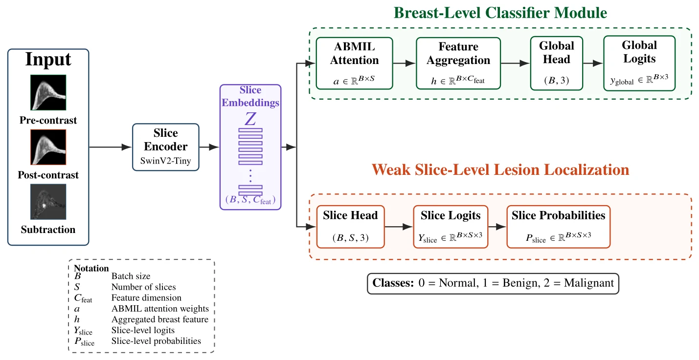
A shared Swin-Tiny encoder feeds two heads: a per-slice lesion probability trained against that Gaussian, and an attention-pooled three-class breast-level classifier. One joint loss, both objectives.
- 01Pre / post / subtraction
- 02Swin-Tiny slice encoder
- 03Slice embeddings
- 04Gaussian slice target
- 05ABMIL pooling
- 06Normal / benign / malignant
03 / WHAT WE TESTED
A multicentre challenge cohort, against progressively stronger baselines.
500 DCE breast MRI cases from multiple European institutions in the ODELIA Breast MRI Challenge dataset, split at breast level with class and institutional balance preserved. Roughly 67% normal, 13% benign, 20% malignant.
Baselines were built up in steps: a Medical Slice Transformer, then the breast-level classifier alone with cross-entropy, then with focal loss, then with slice-level guidance added.
04 / MAIN RESULT
The slice signal helps the diagnosis, not just the picture.
Each component adds something, but the slice-level branch is what pushes AUC past 0.86 and the ODELIA score past 0.70. Against the Medical Slice Transformer that is +7.2 pp AUC and +14.0 pp ODELIA score.
The per-class breakdown shows the gain concentrated on benign cases — the class that is hardest to separate and the one where weak spatial guidance has the most to add.
| Model | Test AUC | ODELIA score |
|---|---|---|
| Medical Slice Transformer (CE) | 0.7968 (0.019) | 0.5695 (0.026) |
| Breast-level classifier only (CE) | 0.8147 (0.007) | 0.5899 (0.025) |
| Breast-level classifier (focal) | 0.8447 (0.002) | 0.6629 (0.010) |
| BE-WISE (CE) | 0.8580 (0.002) | 0.6810 (0.04) |
| BE-WISE (focal) | 0.8683 (0.014) | 0.7098 (0.02) |
ODELIA score = mean of AUC, sensitivity at 90% specificity and specificity at 90% sensitivity. Significance: p < 0.001 (AUC), p < 0.01 (ODELIA).
05 / WHAT THE MODEL SHOWS
The per-slice curve is the explanation.
For correct cases the predicted probability peaks at or next to the slice the radiologist marked. For misclassified cases the curve is flatter or shifted — which is itself informative, because it shows where the model was looking when it got the answer wrong.
Normal exams stay uniformly low, so the branch is not simply firing on every volume.
06 / WHY IT MATTERS
Cheap supervision, inspectable output.
One slice per exam is an annotation budget a clinical partner can actually agree to, and the result is a model whose evidence a radiologist can check in one glance rather than trust on faith.
It also gives a human-in-the-loop path: verify, correct or discard the suggested slice, and feed that back.
LIMITATION
What this does not show
One marked slice is approximate guidance, not lesion delineation — it will not capture multifocal disease or non-mass enhancement well. Annotation variability and lesion-size effects were not analysed, and no reader study was run.
READ IT YOURSELF
Paper · DOI · Code · Citation
View original abstract + citation
Abstract
Breast MRI offers superior soft-tissue contrast and lesion conspicuity compared with mammography, but deep learning on volumetric multi-sequence exams normally requires fine-grained lesion or voxel annotations. BE-WISE asks whether a single radiologist-marked slice per examination is enough supervision for both diagnosis and localization. Pre-contrast, post-contrast and subtraction volumes are stacked as three channels and encoded slice-wise by a shared Swin-Tiny Transformer. A slice-level lesion predictor is trained against a Gaussian target centred on the annotated slice, while an attention-based multiple-instance-learning branch aggregates the same embeddings into a three-class breast-level diagnosis. On the multicenter ODELIA Breast MRI dataset the joint objective improves both breast-level discrimination and the spatial plausibility of the slice-level evidence.
Citation
A. Bhandary Panambur, T.-T. Nguyen, S. Bayer, A. Maier. BE-WISE: Breast MRI Evaluation with Weakly-informed Slice-level Explanation. Bildverarbeitung für die Medizin (BVM) 2026, pp. 10–17. doi:10.1007/978-3-658-51100-5_2